INTERACTIVE EXPLANATIONHow does a reflex help your body react?
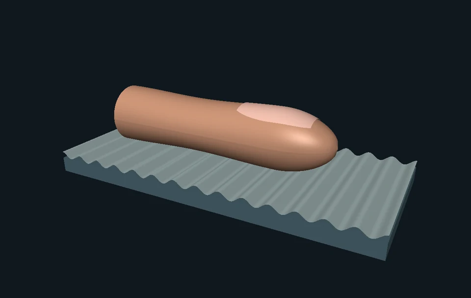
Tip a virtual platform under a real 3D ankle. Discover why one muscle gets more drive while its partner gets less, follow the spinal circuit, and change one connection to see the difference.
Enable JavaScript to change the conditions and run the interactive experiment.
Make a discovery
Your nervous system coordinates muscles. In one stretch-response pathway, it increases drive to a stretched muscle and reduces drive to its opposing partner. That reduction is called inhibition. Try removing the connection and compare both outputs.
- Trace sensory input into the cord and motor output toward a muscle.
- Explain why an inhibitory connection can help coordinate opposing muscles.
- Distinguish a muscle-stretch input from a skin-evoked withdrawal response.
- Recognize that fast responses and crossed responses depend on the pathway and task.
- Read a timing claim by identifying exactly what interval was measured.
- Explain why this model cannot measure someone’s pain, strength or balance.
Make a prediction
Keep the same stretch. Remove only the inhibitory connection. Which output changes in our completed model?
- The opposing muscle keeps more drive
- The calf must receive less drive
- The person’s pain must increase
Read the explanation
The direct branch still adds the same calf drive. Removing the selected inhibitory branch leaves the opposing output at its background value. That is a circuit comparison, not a pain or balance prediction.
Understand it
Where does a stretch response start?
A muscle spindle is a sensory structure inside a muscle. When the calf muscle–tendon arrangement is lengthened by the platform, spindle input can change. Our enlarged spindle is a locator illustration. Its brightness represents a selected teaching stage, not measured nerve firing.
Why show two muscles?
Soleus, at the back of the lower leg, contributes to pointing the toes downward: plantarflexion. Tibialis anterior, in front, contributes to lifting them: dorsiflexion. These are opposing actions at the ankle. Many other muscles, body loads and control pathways also contribute during real movement.
What is a reflex arc?
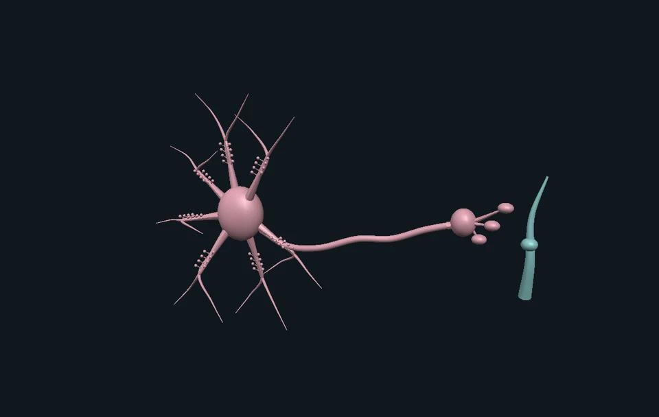
It is a way to trace an input through a nervous-system pathway to an output. The selected stretch example begins with sensory input, includes spinal connections and ends with motor output. The phrase does not mean that every reflex has one identical circuit or that the brain is always absent.
What is reciprocal inhibition?
A sensory branch excites the motor pool of the stretched muscle. Another branch excites an inhibitory interneuron. That neuron reduces drive in the opposing motor pool. The inhibition is a connection within the nervous system; it is not a special inhibitory motor nerve switching off the muscle itself.
Does the signal have to visit the brain first?
The direct spinal branch shown here need not wait for a conscious decision. But the brain can influence spinal circuits, and some fast feedback responses include cortical processing. Automatic does not mean the nervous system above the spinal cord is uninvolved.
Is pulling away from something the same circuit?
Skin-evoked withdrawal starts with a different input and recruits other pathways. Human experiments show that stimulation at different sole locations can produce different muscle-response patterns. A stretch pathway is useful to understand, but it is not a complete diagram of every protective movement.
Look closer at the science
A sensory route and a motor route
Sensory fibers enter through dorsal, or posterior, roots. The sensory cell body is in a dorsal-root ganglion outside the cord; it is not an extra compulsory relay between two neurons. Motor axons leave through ventral, or anterior, roots. Our detached diagram has dorsal at the top and ventral at the bottom. It does not align spinal segments with vertebral levels.
Count central synapses carefully
The selected direct Ia afferent-to-motor-neuron branch has one central synapse. The reciprocal branch includes an inhibitory interneuron, giving two central synapses. Both include additional events elsewhere, including the neuromuscular junction. Neither count is a complete description of every stretch-related response.
The small model we actually calculate
Let u be authored stretch input from 0 to 1, b background drive, and g be 1 with the inhibitory connection or 0 with it removed. At the completed teaching stage: calf drive = clamp(b + 0.5u); opposing drive = clamp(b − 0.3gu). Clamp keeps outputs in [0,1]. The gains 0.5 and 0.3 are chosen for a readable comparison. They are not fitted nerve, EMG, force or movement measurements. During playback we reveal the result in stages, on an illustrative clock.
An extra relay is not the whole response time
Crone and colleagues studied reciprocal ankle pathways in 60 humans in 1987 using conditioned H reflexes. Assuming symmetric central connections, they inferred about 1 ms of additional central delay relative to monosynaptic Ia excitation. That comparison does not measure the whole time from an everyday stimulus to movement, and it says nothing about when someone becomes aware of pain.
Fast feedback can include the cortex
Pruszynski and colleagues combined human and rhesus-monkey experiments in 2011 to investigate rapid multijoint feedback and motor-cortex contributions. This evidence expands the simple spinal picture. It does not make our ankle illustration a model of every arm response or assign one delay to all people.
The other side changes with the task
Suzuki and colleagues compared crossed cutaneous effects on a soleus H reflex in humans. They found facilitation while standing at selected conditioning-to-test intervals, but suppression during early stance in walking. The measured endpoint was electrically probed reflex excitability, not whole-leg force. Gervasio and colleagues separately studied crossed responses during walking and distinguished measured data from modeled afferent activity.
Reflex measures are not a pain meter
Nociception concerns neural processing of potentially tissue-damaging input. Pain is a personal sensory and emotional experience. Experimental reflex measures and reported pain can dissociate. This lesson neither assigns a pain score nor tests a learner’s neurological function.
Where this is used
Staying coordinated when the ground changes
Body motion changes sensory input. Spinal pathways, brain pathways and muscle mechanics together contribute to responses. The ankle reveals a coordination principle, while actual balance needs much more than two output bars.
Why a robot needs feedback too
A robot controller can compare a desired condition with sensor input and adjust its outputs. This is a useful engineering analogy for feedback. A two-motor program is not a biological reflex circuit, and muscles have their own changing mechanical properties.
What scientists and clinicians measure
An electrical muscle recording, a joint movement and a person’s report answer different questions. Research and clinical tests need controlled procedures and interpretation; watching or moving this model is not such a test.
Try it yourself: Build a paper coordination circuit
Supplies
- Paper and a pencil
- Six large paper squares, or six drawn counters
- A second sheet to cover one connection
- Give both outputs a starting level
Draw two boxes: “toes down” and “toes up.” Put or draw two counters in each. Counters represent model drive. They do not measure muscle force.
- Give the input two routes
Draw a “stretch input” card. Connect it directly to the toes-down box. Draw a second route through a card labeled “inhibitory relay,” ending at the toes-up box.
- Make your prediction
Our rules are: the direct route adds one counter; the inhibitory route removes one. Before moving anything, predict both final counts and explain the reason.
- Follow both connections
Activate the input card. Add one counter to toes down and remove one from toes up. You now have three and one. Compare both with the starting counts.
- Cover just the inhibitory connection
Reset to two counters in each box. Cover the inhibitory route and repeat the same input. Toes down reaches three again; toes up stays at two. Which connection explains the difference?
- Explain what your paper cannot tell you
Explain excitation and inhibition to a partner. The counters show the signs of a small circuit. They cannot predict real movement, response time, strength or pain. Those require different evidence.
Can reducing one output change the result without changing the other?
This is a paper model, not a body test. An adult can prepare large squares, or simply draw and cross out counters. No tapping tendons, painful contact, electrical stimulation or sudden loads. No movement is required.
Check your understanding
Which route carries the motor output toward a muscle in this spinal example?
- Dorsal sensory root
- Ventral motor root
- Central canal
Answer and explanation
Ventral motor root Motor axons leave through ventral roots. Dorsal roots carry sensory input; the central canal is not the outgoing muscle-signal route. See FIPAT in the source record.
What does the inhibitory interneuron do in our selected branch?
- Sends an inhibitory motor nerve directly to muscle
- Adds a compulsory stop at the brain
- Reduces drive in the opposing motor pool
Answer and explanation
Reduces drive in the opposing motor pool The sensory branch excites this interneuron, whose output inhibits the target motor pool. The skeletal motor output itself remains excitatory at muscle. Day et al. supports the reciprocal-pathway distinction.
Removing inhibition leaves more opposing drive in our model. What follows?
- The model has more opposing drive
- Every real person must fall
- Their pain must increase
Answer and explanation
The model has more opposing drive Only the first claim is calculated here. The platform sets foot pose; no whole-body balance or pain is predicted.
What changes when we move from a stretch example to skin-evoked withdrawal?
- Skin turns into muscle
- The input and recruited circuit change
- All interneurons disappear
Answer and explanation
The input and recruited circuit change A muscle spindle supplies the main example. Skin input recruits other pathways and location-dependent patterns, as examined by Andersen et al.
Does a fast automatic response prove that the brain is uninvolved?
- Yes, always
- Only if both legs move
- No; some fast feedback includes cortical processing
Answer and explanation
No; some fast feedback includes cortical processing A direct spinal branch need not wait for a decision. Other fast feedback pathways can include cortex, as tested by Pruszynski et al.
Must the opposite leg always show the same crossed response?
- Yes
- No; task and pathway matter
- Only in children
Answer and explanation
No; task and pathway matter Suzuki et al. found different crossed effects on a tested H reflex during standing and early stance in walking. That is a context comparison, not a universal rule for leg force.
A study infers one extra millisecond of central delay. Did it measure a one-millisecond stimulus-to-movement time?
- Yes
- Yes, but only for pain
- No; those are different intervals
Answer and explanation
No; those are different intervals Crone et al. compared selected central pathway delays under an assumption. Sensory travel, motor travel and movement are not all included in that extra central interval.
Can a larger reflex automatically tell us how much pain someone experiences?
- No
- Yes, exactly
- Only if playback is slow
Answer and explanation
No Pain is personal. Reflex measures and reported pain can dissociate; the IASP definition and Edwards et al. support keeping those measures distinct.
Sources and model limits
- Anatomy comes from selected BodyParts3D 4.0 right-sided muscles, tendon and bones. The platform, spindle locator, articulation and functional circuit are authored illustrations, not an observed reflex in motion.
- The ankle uses an approximate single pivot and a smooth deformation of selected soft tissues. The platform prescribes the foot pose. Outputs do not solve joint torque, balance, muscle force, tendon strain or realistic spindle dynamics.
- Model drive is a normalized teaching quantity. The gains and playback stages are authored; there are no physiological milliseconds, Hz, EMG or force units attached to them. Removing inhibition is a counterfactual circuit comparison, not a diagnosis.
- The circuit selects one Ia stretch-response branch. It omits many neurons, inputs, presynaptic regulation, descending influences and task-dependent dynamics. Inhibition is shown at the opposing motor pool.
- Withdrawal and crossed-response evidence concerns different inputs and experiments. The standing/walking comparison is qualitative evidence about an H reflex, not a gait simulator.
- The scientific figure is a static diagram of a research setup. It is not a photograph, a result graph or a home experiment. Source geometry and figure attribution are downloadable. Independent subject and learner review of the finished lesson remains pending.
Human ankle pathways support reciprocal Ia inhibition
Crone et al. studied 60 humans using conditioned soleus and tibialis-anterior H reflexes. The extra central delay was inferred under a symmetry assumption; our drive gains are not taken from their data.
Crone et al. · Journal of Physiology, 1987An inhibitory interneuron explains the selected reciprocal branch
Original human forearm conditioning experiments; corroborating pathway evidence, not ankle-specific simulation constants.
Day et al. · Journal of Physiology, 1984Human muscle spindle responses depend on mechanical conditions
Original recordings from human muscle spindle afferents during vibration, length and movement conditions. The lesson does not reproduce their firing rates.
Burke et al. · human spindle recordings, 1976Skin-evoked withdrawal patterns depend on input location
Original human foot-sole electrical-stimulation study. Not every withdrawal response is one universal activation pattern.
Andersen et al. · human leg withdrawal, 1999Some fast feedback involves motor cortex
Primary human and rhesus-monkey experiments on multijoint feedback control. Distinguish these experimental tasks from the modeled ankle.
Pruszynski et al. · Nature, 2011Crossed effects differ during standing and walking
Human conditioned soleus H-reflex measurements. The 100–130 ms intervals are between conditioning and test stimuli, not natural movement onset delays.
Suzuki et al. · Experimental Brain Research, 2014Walking research separates measurements from estimated neural activity
Gervasio et al. 2017, PLOS ONE 12(1):e0168557. Complete Figure 3 documents the setup and is reused under CC BY 4.0. This paper is separate from Suzuki’s standing/walking comparison.
Gervasio et al. · PLOS ONE, 2017Inhibition has a long experimental history in coordination
Sherrington’s own 1932 Nobel lecture. Historical animal experiments are not direct human measurements or the origin of our authored numerical gains.
Sherrington · original Nobel lecture, 1932Dorsal and ventral roots have distinct anatomical identities
FIPAT Terminologia Neuroanatomica. Terminology guides the detached functional diagram; it does not validate authored geometry.
FIPAT · Terminologia NeuroanatomicaThe 3D anatomy is from the dataset owner’s source meshes
BodyParts3D, © The Database Center for Life Science licensed under CC Attribution 4.0 International. Current owner license updated February 2025; source selections and changes are recorded in the manifest.
DBCLS · BodyParts3D license and attributionPain is not inferred solely from sensory-neuron activity
IASP’s definition distinguishes personal pain experience from nociception. No pain prediction or self-test is offered.
IASP · revised definition of painReflex measures and pain ratings can dissociate
Edwards et al. original human study of task-related arousal, reflex threshold and pain ratings. The lesson uses the distinction, not a personal calibration.
Edwards et al. · Journal of Psychophysiology, 2006Independent subject review is pending.
Read the sources and model assumptions