INTERACTIVE EXPLANATIONHow does a nerve cell send a message?
Nudge a membrane, discover when a spike appears, test its recovery, and cross a chemical synapse. Then inspect a real potassium-channel structure.
Enable JavaScript to change the conditions and run the interactive experiment.
Make a discovery
A nerve impulse is a changing pattern of membrane voltage. Ions cross nearby membrane regions; they do not carry a parcel the full length of the axon. At a chemical synapse, a different process releases molecules that can change the next cell’s input.
- Locate dendrites, cell body, axon and a separate receiving cell.
- Distinguish a small graded voltage response from an action potential.
- Use sodium and potassium conductances to explain rising, falling and recovery phases.
- Compare two second pulses without inventing a universal recovery timer.
- Order calcium entry, vesicle fusion, transmitter release and receptor binding.
- Distinguish a measured molecular structure, an illustration and a quantitative membrane model.
Make a prediction
Will doubling a short input pulse make a spike twice as tall?
- Yes: a nerve cell is a linear amplifier
- Not in this membrane model
- It must produce exactly two spikes
Read the explanation
Try 20 and 40 μA/cm² for 1 ms. Both produce one spike with similar, not identical, peaks. The spike emerges from voltage-dependent conductances.
Understand it
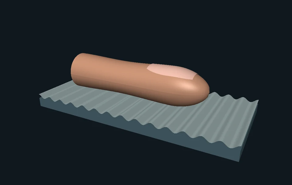
How does your skin feel a touch?
When something presses on your skin, specialized sensors can start electrical messages in sensory nerves. Your nervous system uses these messages to help you feel a touch. Signals about potentially harmful stimuli are part of a more complex process: pain is an experience, not just an electrical pulse. This lesson explores one foundation: how a nerve cell sends a message. Its classic squid-membrane experiment does not simulate skin receptors or predict what a person feels.
Many places to receive input
Dendrites and the cell body integrate inputs from other cells. Branching morphology and membrane properties affect the result. The illustrated nucleus belongs to the cell’s internal machinery; it is not the point that fires an electrical spark.
Voltage is a difference across a membrane
Here membrane voltage means inside minus outside. A less negative value is a depolarization. The pulse bench injects a specified current density into a uniform membrane patch and computes its response. Small inputs can produce a graded change that settles back toward rest.
A regenerative response can appear
Voltage-dependent sodium activation can increase inward sodium current, which further depolarizes the membrane. This feedback can produce an action potential. The bench solves continuous voltage and gating equations: it has no separate command to fire at −55 mV or to reset the voltage afterward.
The currents overlap
Sodium availability decreases through inactivation while potassium conductance rises with a delay. Outward current helps bring voltage down; elevated potassium conductance can contribute to an undershoot. The sodium and potassium processes overlap rather than taking perfectly separate turns.
Recovery changes the next response
After a spike, the membrane’s gating state differs from its initial state. In one tested comparison, a second 20 μA/cm² pulse at 10 ms onset spacing fails to produce a second spike, while 40 μA/cm² succeeds. This demonstrates relative recovery in the chosen model and protocol, not a universal human recovery duration.
A chemical synapse changes the message format
Presynaptic depolarization can open calcium channels. Local calcium entry helps trigger vesicle fusion and transmitter release. Molecules diffuse to receptors across a separate cleft. The receiving cell gets a graded input; its response depends on receptors, other inputs and its current state. Electrical synapses provide another form of connection, outside this animation.
Look closer at the science
A specified historical membrane experiment
The Hodgkin–Huxley bench represents one uniform squid giant-axon membrane compartment at 6.3°C. It uses the modern NEURON voltage convention, not the original paper’s resting-relative sign convention. Capacitance is 1 μF/cm²; maximum Na/K/leak conductances are 120/36/0.3 mS/cm² and reversal potentials are +50/−77/−54.3 mV.
The voltage equation
C dV/dt = Iapp − INa − IK − IL. INa = 120 m³h(V − 50), IK = 36 n⁴(V + 77), and IL = 0.3(V + 54.3). Positive ionic current is outward; positive applied current depolarizes. m is sodium activation, h is sodium availability against inactivation, and n is potassium activation. Gating variables are continuous model states, not literal moving door counts.
Equilibrium and numerical time
The exact zero-current equilibrium for these parameters is about −64.97405 mV, with gates initialized at their steady values. All four differential equations use RK4 with steps no larger than 0.005 ms, split at stimulus boundaries. Rendering speed does not change this integration. Reference traces, pulse counts, removable rate singularities and convergence are checked separately.
Counting is not triggering
A spike is counted at an upward 0 mV crossing, with its time interpolated between adjacent samples. This is a detection convention for these experiments, not the voltage at which the biological feedback begins. The solver does not change its equations at that crossing. Stronger input does not impose a proportionally taller spike.
Voltage clamp asks another question
In the optional fixed −20 mV demonstration, voltage is held constant while gates relax analytically from the resting values. Sodium conductance rises then falls; potassium conductance increases more slowly. That is a voltage-clamp response, not a freely evolving action potential. At 1 ms the computed gNa/gK are about 17.404/2.241 mS/cm²; at 5 ms they are about 1.484/13.026.
Ion channels and pumps have different roles
Fast spikes depend on channel currents and capacitance. Na/K pumps help maintain the concentration gradients, exchanging three Na outward for two K inward per cycle; they are not an instantaneous reset after every spike. This HH model holds reversal potentials fixed and has no explicit pump, changing ion concentration or spatial propagation state.
From nanometers to a whole cell
The 2R9R reference preserves a static biological assembly of four engineered Kv1.2–Kv2.1 channel subunits with four beta subunits. It is a rat-derived molecular example, not the molecular identity of the squid model’s potassium current. The synapse uses an illustrative 25 nm cleft, within the 20–30 nm range reported for the cited adult-rat synapses. The cell illustration and membrane icons use separate, unmeasured display scales.
Where this is used
Why a single message can have many outcomes
A chemical input may meet a cell already near a regenerative response, a recently active cell, or different receptor machinery. A neural network depends on these interactions; a single visible spike is not a complete thought.
How researchers ask controlled questions
A current pulse asks how voltage responds to input. A voltage clamp holds voltage and asks how currents change. Switching between the two exposes different parts of the mechanism instead of treating every trace as the same experiment.
What a molecular structure adds
The source-based channel model lets you inspect physical protein organization that the HH equations deliberately compress into conductance variables. Structure, electrical behavior and synaptic communication answer different questions.
Try it yourself: Build a wave that needs time to recover
Supplies
- 12 numbered paper cards
- A pencil
- Four state marks: ready, active, recovery 1, recovery 2
- Four small slips for chemical-synapse events
- Make twelve local cells
Lay the numbered cards in a row. Each starts ready. Record all twelve states in one row of a notebook before changing anything.
- Start a pattern at one end
Mark card 1 active. On the next round, a ready card beside an active card becomes active. Active becomes recovery 1; recovery 1 becomes recovery 2; recovery 2 becomes ready. Update every card from the previous recorded round.
- Watch the recovering tail
Repeat the simultaneous update. The active pattern moves, but the cards do not travel along the row. Try reactivating card 1 after one round and after four rounds; only a ready card can be activated in this toy rule.
- Start in the middle
Restore all cards to ready, activate card 6, and update together again. Two fronts spread away from the start. A recovering region and the place of initiation help determine the pattern’s direction.
- Cross a different kind of connection
Draw a gap after the last card and place a separate receiving card beyond it. Order the slips: calcium entry, vesicle release, transmitter binding, postsynaptic input. Do not pass the active-state mark straight across the gap.
- Compare the analogy with the experiment
The cards use an absolute waiting rule. The membrane bench also lets a stronger input succeed during relative recovery. Explain what the cards show well and one important feature their rules cannot capture.
Can a pattern move while every paper card stays in its own place?
A tabletop analogy with chosen discrete rules. No electrical stimulation or body measurements. Card rounds and dimensions are not physiological milliseconds or nerve lengths.
Check your understanding
What moves along the illustrated axon during a nerve impulse?
- One sodium ion travels from end to end
- A pattern of changing membrane voltage
- A neurotransmitter-filled vesicle crosses the whole axon instantly
Answer and explanation
A pattern of changing membrane voltage Ions cross locally as successive membrane regions change voltage. The moving color represents that pattern, not one traveling ion.
A small pulse raises voltage slightly, then it returns toward rest. What happened?
- A graded response without a detected spike
- The model lost a mandatory spike
- A guaranteed signal reached the second cell
Answer and explanation
A graded response without a detected spike A change in membrane voltage need not become a regenerative spike. Compare the 5 and 20 μA/cm² trials.
Which explanation fits the model’s falling phase?
- The pump instantly puts every ion back
- Outward K current with reduced Na current helps bring voltage down
- The membrane runs out of electrons
Answer and explanation
Outward K current with reduced Na current helps bring voltage down Sodium inactivation and delayed potassium activation overlap. The HH model has no explicit pump state and still generates a falling phase.
At 10 ms spacing, a stronger second pulse succeeds when the weaker one fails. What does this demonstrate?
- A universal human recovery time of 10 ms
- Relative recovery in this tested protocol
- An absolute inability to generate another spike at that time
Answer and explanation
Relative recovery in this tested protocol The membrane’s state changes the input needed for a second response. These results do not establish one refractory duration for all cells.
Which chemical-synapse order is correct?
- Receptor binding → calcium entry → depolarization
- Depolarization → calcium entry → vesicle release → receptor binding
- An intact vesicle jumps into the receiving cell
Answer and explanation
Depolarization → calcium entry → vesicle release → receptor binding Local calcium entry helps trigger vesicle fusion. Transmitter molecules then reach receptors across the cleft.
Does one released chemical message guarantee one spike in the next cell?
- Yes, always one
- No; receptors, other inputs and cell state matter
- Yes, always two
Answer and explanation
No; receptors, other inputs and cell state matter The recipient receives a postsynaptic input. This qualitative synapse does not calculate a guaranteed output spike.
The 100 ms trial gives 7 spikes at 10 μA/cm² and 9 at 20. What can you conclude?
- Doubling input doubled the spike count
- The tested counts changed without doubling
- All neuron spike heights doubled
Answer and explanation
The tested counts changed without doubling Read the actual trials. Count, timing and some waveform details can change. Neither height nor count is imposed as a linear multiple of input.
Can the uniform squid-patch trace and illustrated axon length give human conduction velocity?
- Yes, divide the picture’s length by playback time
- No; this model has no spatial propagation or human calibration
- Yes, if you use imperial units
Answer and explanation
No; this model has no spatial propagation or human calibration You would need a spatial model or measured distance/time data. Illustration pacing and a one-compartment voltage equation cannot provide that result.
Sources and model limits
- The HH bench is one deterministic uniform squid membrane patch at 6.3°C. It cannot calculate human conduction velocity, thoughts, diagnoses, learning or disease.
- The whole-cell geometry and propagation highlight are original teaching illustrations. Displayed axon length and playback speed are not measured physiological values.
- The model fixes ion gradients, temperature and channel parameters. It omits stochastic channel openings, pumps, axonal cable currents, synaptic kinetics and changing concentrations.
- Chemical-synapse events and the calcium-closure comparison are qualitative. They do not calculate release probability, diffusion constants, postsynaptic spike timing or a drug effect.
- The 2R9R asset is a smoothed protein-coordinate envelope from biological assembly 1. Lipids, ions and small ligands are omitted. Mesh smoothing is not experimental resolution or a measured gating motion.
- The paper activity has authored discrete recovery rules. Its rounds do not measure the refractory period of the HH model or a person.
Human evidence that PIEZO2 contributes to touch and body-position sensing
Two participants with loss-of-function variants; sensory effects differed by test. Context for sensory signaling, not validation of the squid-membrane simulation.
Chesler et al. · Human mechanosensation, NEJM 2016Pain is an experience distinct from nociception
The revised definition distinguishes pain from activity in sensory neurons. A voltage trace alone does not measure what a person feels.
IASP · Revised pain definitionVoltage-dependent sodium and potassium conductances generate membrane responses
Original uniform-membrane equations, Table 3, excitation and recovery analysis; distinguish uniform from spatially propagated responses.
Hodgkin & Huxley · Journal of Physiology, 1952Exact modern-convention rate and current equations
Pinned hh.mod revision 8202fe7. Our original solver uses direct rate functions, specified reversal potentials and RK4; it does not copy the optional voltage lookup table or use NEURON as a runtime.
NEURON · official HH mechanism sourceMembrane capacitance and Na/K reversal values used in the numerical convention
Chapter 6’s section properties and mechanism examples. Values are converted to mS/cm² and μA/cm² in the lesson.
The NEURON Book · chapter 6Neuron structure, chemical/electrical synapses and graded postsynaptic input
Authors’ online textbook, sections 1.1 and 1.2. Original illustrations are authored for Brytalearn; textbook figures are not reproduced.
Gerstner et al. · Neuronal DynamicsAxonal initiation in a specified mammalian preparation
Rat layer-5 pyramidal-cell research. Supports separating axonal initiation from the nucleus without universalizing every detail to all neurons.
Stuart, Schiller & Sakmann · 1997Sodium/potassium gradients and pump exchange
Institutional explanation of ion signaling and three-Na/two-K pump exchange; pump maintenance is distinct from the fast model waveform.
NIH NIGMS · sodium and nerve signalingVesicle release, receptor binding and transmitter clearance
Institutional explanation of chemical communication. Receiver effects depend on the receiving cell; a released vesicle does not itself travel intact across the gap.
NIH NIGMS · What Is a Neurotransmitter?Presynaptic calcium entry triggers release in an experimentally studied chemical synapse
Rat calyx-type preparation; no preparation-specific channel count is assigned universally in this animation.
Borst & Sakmann · Nature, 1996The illustrative cleft dimension has a preparation-specific comparison
Electron tomography study of adult-rat cortical/hippocampal axospinous synapses, synaptic-cleft section. Reported 20–30 nm range; 25 nm here is an illustrative choice.
Burette et al. · Journal of Comparative Neurology, 2012A real potassium-channel molecular assembly
PDB 2R9R biological assembly 1, A4B4, entry revision 1.2. Original structure by Long et al., Nature 2007, DOI 10.1038/nature06265. The 2.4 Å value is experimental resolution.
RCSB PDB · 2R9RDeposited coordinate data may be reused with their provenance
PDB archive data are CC0. Local processed protein envelopes preserve chain/entity identities, coordinate transform and checksums; credit Long et al. and the archive.
RCSB PDB · data policiesIndependent subject review is pending.
Read the sources and model assumptions