Start with normal function
Sebaceous glands make an oily material called sebum. A short duct joins the upper hair follicle, and the normal route reaches the surface. The surface pore is an exit, not the source of the oil.
Open a connected follicle, follow its normal oil route, and compare structures hidden beneath the surface. Read real human microscopy and discover why acne is more than a story about dirt.
Enable JavaScript to change the conditions and run the interactive experiment.
A skin opening is only the part we can see from above. Underneath, a gland connects to a hair follicle. Oil, shed cells, resident microbes and local responses all belong to the story. Different structures can look similar at the surface; a surface color is not a hygiene score or a diagnosis.
Which statement correctly separates an opening from its contents?
Open describes exposure at the surface. It does not establish an empty channel; similar surface colors can hide different structures.
Sebaceous glands make an oily material called sebum. A short duct joins the upper hair follicle, and the normal route reaches the surface. The surface pore is an exit, not the source of the oil.
Keratinocytes line the follicle. They are cells; keratin refers to structural proteins. Retained keratin-containing material inside a follicle is different from dirt that arrived from outside.
In a closed comedo, retained material lies beneath a cover. In an open comedo, a plug reaches an exposed opening. Open does not mean the passage is empty.
A blackhead is an exposed comedonal plug. Its dark appearance is not evidence that someone did not wash enough. This model does not turn surface color into a bacterial count, severity score or oxidation clock.
A closed comedo is not defined by being full of pus. A pustule is a pus-containing inflammatory structure. The cutaway and material key distinguish them even when their surface colors look similar.
Cutibacterium acnes can occur on skin with and without acne. Its presence alone does not establish a diagnosis. Microbial strains, their surroundings, skin cells and local responses can matter together.
Researchers have detected inflammatory changes around follicles before obvious surface changes. These five authored cases therefore compare structures. They are not a guaranteed sequence, a disease timer or a prediction of what a person’s skin will do.
The fluorescence panels show fixed, stained human tissue. Researchers chose labels that create the colors. The same color can represent different labels in different panels, so each image needs its own key.
A hair follicle and its associated sebaceous gland form a connected anatomical unit. The gland has lobules and joins the upper follicle through a duct. Our original geometry makes those connections inspectable; it is not a patient scan or a universal set of dimensions.
The follicle lining, hair shaft, gland, duct and surface opening have different roles. The infundibulum is the upper follicular region leading toward the surface. The model keeps the lining distinct from retained material.
Durgin and colleagues’ human microscopy shows gland lobules, a thin peripheral basal-cell layer and differentiated sebocytes. The model’s colors and cell-like surface patterns help identify structures; their number and geometry do not measure those source cells.
Hormonal changes around puberty can affect sebaceous-gland activity. Age, inherited factors and local biology also matter. No validated personal prediction follows from changing an arbitrary oil or hormone value, so this lesson does not provide those controls.
Comedones involve enlarged or plugged follicles. Covered material and exposed material differ in their connection to the surface. The amount, shape and depth in this model are authored examples rather than clinical cutoffs.
Jeremy and colleagues examined cellular, vascular and proliferative markers in biopsied follicles. Their findings support early inflammatory involvement, including changes in clinically uninvolved skin. The original abstract does not supply a universal timetable for a particular person.
Fitz-Gibbon and colleagues sampled nasal pilosebaceous units from 49 acne participants and 52 controls. The relative abundance of the species was similar while strain-population structures differed. This site-specific association study does not define a universal healthy/acne threshold or prove that any detected strain alone caused a lesion.
Kim and colleagues used cell experiments and tissue localization to investigate TLR2-related cytokine responses to P. acnes, the older species name. Such experiments support a mechanism under their conditions. They do not mean all resident microbes are invaders or one pathway explains every case.
In Durgin Figure 1A, KRT5 is green and KRT79 red. In Figure 1B, KRT5 is red and PPARG green; the overview also shows DAPI in blue. These are research labels. Red does not automatically identify inflammation, and segmentation dots in later panels are not bacteria.
Moving our plane can make it miss a gland that still exists elsewhere in the model. Real tissue sections likewise reveal a particular sampled preparation. The static source photograph cannot be rotated into a newly measured 3D volume.
The paper’s caption reports 50 μm scale bars. This lesson does not measure the A/B crops without identifying a visible bar. Separately, 50 μm equals 0.05 mm, so twenty such lengths equal 1 mm. The unit conversion does not establish a gland’s dimensions.
The spatial-transcriptomics study analyzed 24 sections from 11 donors using a targeted gene panel. Those are not 24 independent people, and gene-expression measurements are not direct measurements of oil output. Source evidence informs the anatomy without becoming a fabricated flow simulation.
Trace the connections and identify what the drawing leaves out. A surface mark and its underlying structure are different observations.
Ask how the image was made, which labels create its colors, and whether it is a section or a volume. Check the panel key before interpreting a color.
Use anatomical terms and avoid treating appearance as a measure of cleanliness, effort or character.
Fold a sheet into an L shape. Label the horizontal face surface and the upright face view inside. Draw a small surface opening and a channel below it.
Draw a lobulated gland beside the upper channel, attached by a short duct. Add a fine hair and label gland, duct, follicle and opening using the reference sheet.
Trace from the gland through the duct and follicle to the surface with a finger on the paper. Explain why the opening is the exit rather than the source.
On the second sheet, make two folded comparisons: retained material reaching the surface and retained material beneath a cover. Use different hatching for the lining and retained material. No real oil or powder is needed.
Show only the surface view. Ask which facts can be observed and which require looking inside. Unfold and compare. These are paper anatomy examples, not a guide for identifying someone’s skin.
Draw a 5 cm line. Label it 50 μm at an invented 1,000× drawing scale. Since 50 μm = 0.05 mm, multiplying by 1,000 gives 50 mm = 5 cm. This one reference line does not put the whole freehand drawing to scale.
What became visible when you unfolded the paper, and what can this model still not tell you?
A paper anatomy investigation only. Do not squeeze, extract, culture or apply anything to skin. The drawing does not establish what caused a person’s acne.
The sebaceous gland The gland connects through its short duct to the upper follicle.
Open describes a plug exposed at the surface. The surface relationship and the retained contents are different facts.
It is an exposed plug, not a hygiene score. A dark comedonal plug is not evidence that someone did not wash enough.
No: a covered plug and a pus-containing inflammatory cavity differ. Their internal structures and contents differ.
No: the species can occur in people without acne. The original comparison found this species in both participant groups, with differences at finer levels.
Cases vary, and inflammation can begin before it is visible. Morphology, mechanism and observed timing are different kinds of evidence.
No: read that panel’s research-label key. Red labels KRT79 in panel A and KRT5 in panel B.
The display enlarges; the real length remains 0.05 mm. Micrometers and millimeters differ by a factor of 1,000; zoom changes the display.
Official anatomy and lesion definitions. Basic explanations are qualified by original research; no personal cause or prediction is inferred.
NIAMS · Acne overviewFigure 1 and the primary spatial-transcriptomics study. CC BY 4.0 images are credited separately; expression findings do not supply sebum flow rates.
Durgin and colleagues · JCI Insight 2026Panel A labels KRT5 green/KRT79 red; panel B changes the key. Complete original and documented panel derivatives are available locally.
Durgin · Original Figure 1 and captionPrimary nasal sampling study of 49 acne participants and 52 controls. Strain associations and relative abundance are not a universal diagnosis rule.
Fitz-Gibbon and colleagues · Strain populationsOriginal abstract read; no uninspected histology images or effect sizes are claimed. Supports avoiding an obligatory inflammation-last timeline.
Jeremy and colleagues · Lesion initiationPrimary cell/tissue study. Mechanism evidence is distinct from a personal treatment recommendation or a claim that every microbe is harmful.
Kim and colleagues · TLR2 investigationTen participants, selected small skin areas and weekly observations over six weeks. Sampling and optical markers limit temporal interpretation; images are not redistributed.
Manfredini and colleagues · Longitudinal imagingOfficial guidance for personal concerns. The lesson provides no medication menu or treatment protocol.
NIAMS · Personal care and professional guidanceIndependent subject review is pending.
Read the sources and model assumptions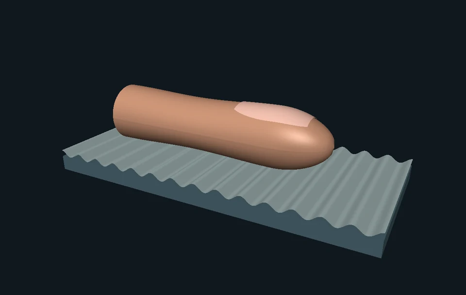 Interactive exploration
Slide a texture beneath a finger. See the contact change, compare a moving touch with a steady one, and zoom from skin to a real sensing protein. Discover how touch differs from pain.
 Interactive exploration
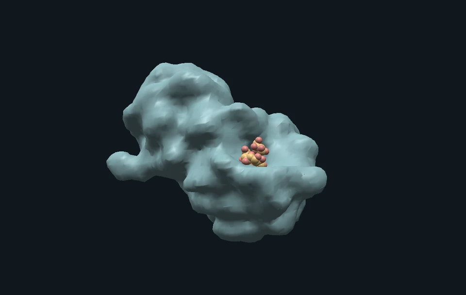
Match two kinds of fiber to the microbes that can use them. Look through a bacterial surface, inspect a real enzyme holding a sugar chain, and sort the different kinds of products that microbes can make.
 Interactive exploration
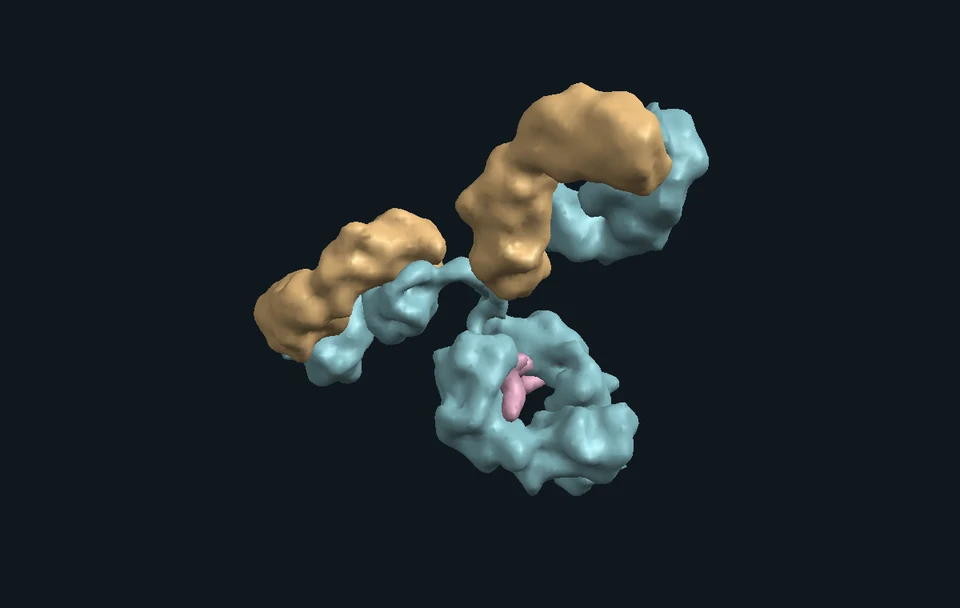
Explore how the immune system responds to an infection. Follow immune cells from skin to lymph node, inspect an antibody structure, and discover what immune memory leaves ready.