INTERACTIVE EXPLANATIONHow does a tiny intruder set a whole response in motion?
Explore how the immune system responds to an infection. Follow immune cells from skin to lymph node, inspect an antibody structure, and discover what immune memory leaves ready.
Enable JavaScript to change the conditions and run the interactive experiment.
Make a discovery
Defense depends on connected jobs: detecting, carrying information, recognizing, cooperating and acting. A memory cell and an antibody are different parts of that system.
- Locate tissue, blood and a draining lymph node in one connected response.
- Distinguish an intact antigen from a peptide displayed with MHC II.
- Explain why a matched B cell and helper T cell cooperate in this protein-antigen example.
- Separate antibody binding, phagocyte uptake and subsequent processing.
- Distinguish memory cells from plasma cells that can keep supplying antibodies.
- Explain what a deposited molecular structure shows and what the simplified response cannot predict.
Make a prediction
If an antibody attaches to a bacterium, has it already destroyed it?
- Yes: attachment means destruction
- No: binding can help a separate effector action
- Yes: the antibody turns into a phagocyte
Read the explanation
The scene separates a matching tag, Fc engagement, engulfment and processing. Antibody proteins and phagocytic cells have different jobs.
Understand it
A familiar starting place: the skin
Skin is a barrier. In this imagined scene, a small breach leaves a generic bacterium in the tissue beneath it. Resident cells can recognize microbial or damage-associated cues. Their signals can help recruit other cells from a nearby blood vessel. The first response does not have to wait for a newly made antibody.
A message travels to a meeting place
A dendritic cell acquires antigen material and travels along a lymphatic route to a draining lymph node. It displays a processed protein fragment—a peptide—with an MHC II molecule. A compatible helper T cell can recognize that display. Additional activating signals matter too; merely seeing something is not an automatic instruction to respond.
Two readers, two forms of the message
The shown B-cell receptor binds an exposed feature of intact protein A. The shown helper-T receptor recognizes peptide pA together with compatible MHC II. These are related through the same protein, but they are different recognition targets. Try presenting the wrong form in the matching lab.
The right cells cooperate
The B cell captures intact protein, processes it and displays a peptide on its own MHC II. A compatible activated helper can provide linked help. The selected B lineage can then produce secretory descendants called plasma cells, as well as a memory branch. The helper cell does not turn into a plasma cell.
A tag helps another cell act
A plasma cell releases antibodies: proteins, not miniature cells. The antigen-binding arms of a matching antibody can attach to a target. A phagocyte can engage the exposed Fc region with a receptor and engulf the tagged target. This assistance is called opsonization. Binding, engulfment and processing are separate events.
Some preparation can remain
After an earlier response, matching memory B and helper T populations may remain. Some long-lived plasma cells can also keep supplying antibody. Compare memory alone with memory plus existing antibody. Then change to antigen B: retained A-specific preparation does not become a universal match.
Look closer at the science
Scope: one T-dependent protein-antigen response
This qualitative scenario concerns a protein antigen on a generic extracellular bacterium. It omits T-independent antibody responses, infected-cell killing, complement, tolerance mechanisms and detailed helper-cell subsets. The grouped activation/help setting stands for multiple biological prerequisites; it is not one universal molecular switch.
Clonal selection and linked recognition
A repertoire of differently recognizing cells already exists. Here, exact A/B/C labels define an authored compatibility relation. A B receptor recognizes intact antigen, then the cell can process it for peptide–MHC II presentation to a compatible helper. Real affinity, cross-reactivity, HLA variation and the full receptor repertoire are not computed.
Compartments and anatomical scale
Blood carries cells; lymphatics connect tissue with draining nodes. The scene separates these routes, then abbreviates efferent lymph, blood circulation and tissue access for returning antibodies. NIH cell and node illustrations are flat images placed in an authored 3D scene. Added B/T regions are explanatory overlays, not a reconstruction of a measured node. Cell, molecule, vessel and organ scales are independently enlarged.
Recognition is not the whole effector mechanism
Fab regions include the antigen-binding arms. Fc can engage effector mechanisms, including suitable phagocyte receptors. Closing Fc access here disables only the selected antibody-assisted uptake route. It does not disable every innate pathway or imply that all antibodies have the same effector action.
Memory versus continuing secretion
A memory B cell can participate in a recall response. A plasma cell is specialized for secretion. The comparison board records which components are available, without imposing a clinical protection duration, antibody concentration or a fixed number of days to clearance.
A real structure has a specific identity
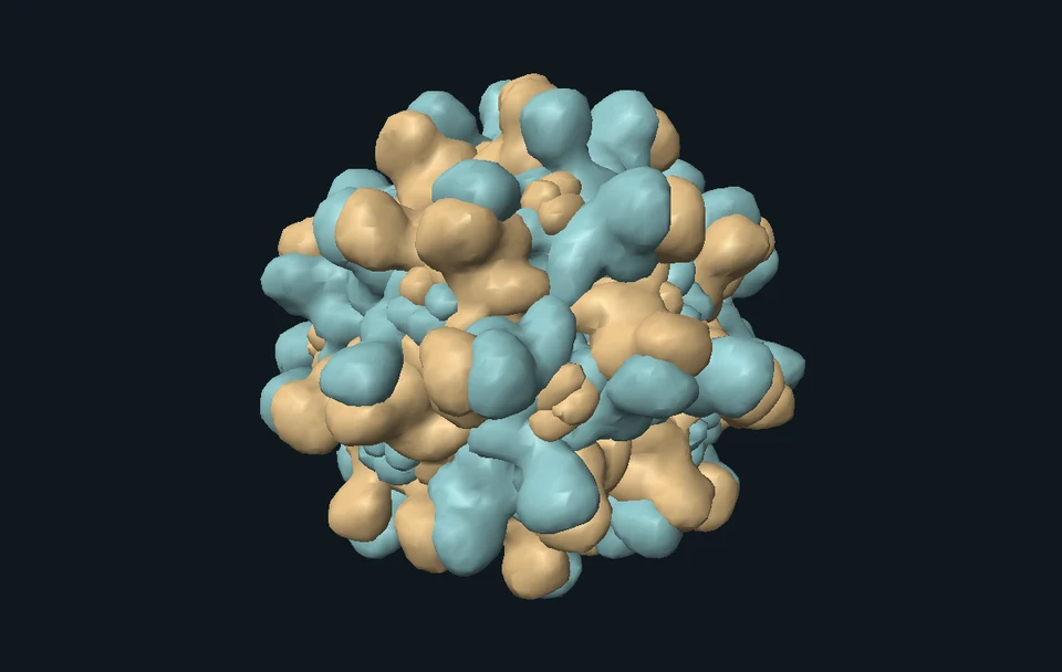
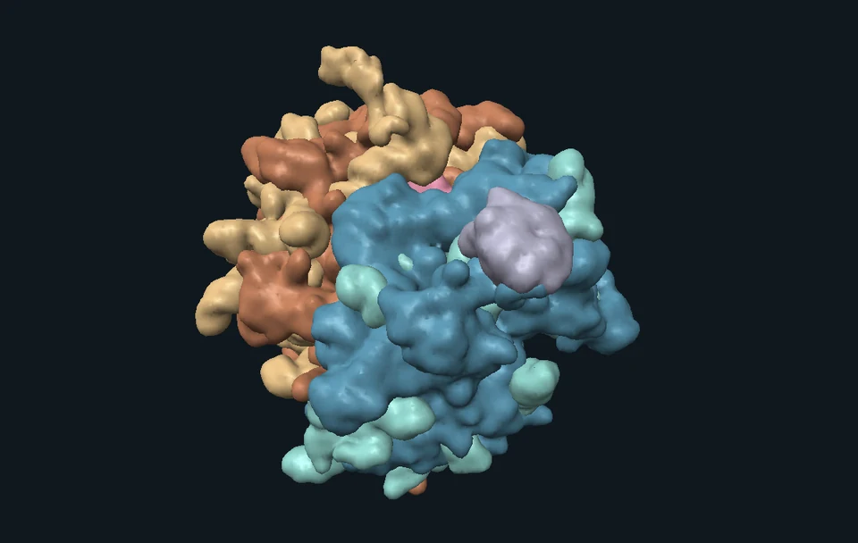
The molecular view uses biological assembly 1 of PDB 1IGT: mouse IgG2a Mab231, studied against a canine lymphoma target. It contains two heavy chains, two light chains and two deposited glycan chains. It is a separate physical example, not an antibody known to bind our fictional protein A. The asymmetric deposited pose is preserved; display separation is not measured molecular motion.
Where this is used
Why a lymph node belongs in the story
Local tissue can communicate with an organized meeting place. Following the route explains why a response involves more than the cells visible beside a skin breach.
Why prior preparation is specific
Memory can change what is available at the start of a later encounter. Vaccination builds on this principle, but each vaccine, antigen and disease needs its own evidence and explanation.
How researchers connect scales
Cell imaging reveals where interactions occur. Molecular structures reveal physical organization. Functional experiments test what changes when a component is altered. None of these alone predicts the course of every infection.
Try it yourself: Build two ways to recognize one message
Supplies
- Paper and a pencil
- Two large protein cards marked A and B
- Six receptor cards: B-A, B-B, B-C, T-A, T-B, T-C
- Small peptide slips pA and pB, plus an MHC II paper frame
- One helper-signal token, two plasma-cell cards and two memory cards
- Prepare before meeting an antigen
Make every receptor card before drawing a protein card. Put tissue and lymph-node labels on opposite sides of the table. The antigen selects among the cards; it does not rewrite them all.
- Try the intact message
Choose protein A. B-A can capture it under our rule. B-B and B-C cannot. Do not put the protein in the MHC frame for this B-receptor step.
- Change the form
Place peptide pA in the MHC II frame on a dendritic-cell area in the node. T-A recognizes that combination. Remove the frame: the peptide alone no longer satisfies this paper T-receptor rule.
- Link capture to help
The B-A cell now displays another pA slip in its own MHC II frame. Bring the compatible activated T-A card and helper-signal token beside it. Only then add a plasma-cell descendant and a separate memory branch.
- Tag, then act
Draw an antibody with two antigen-binding arms and an Fc stem. Match its arms to protein A. Use a separate phagocyte card to engage the stem, engulf the paper target and then mark it processed. Keep these three actions distinct.
- Try again with something different
Keep the A memory cards. First repeat A with memory alone, then add a retained plasma-cell card that supplies existing anti-A antibody. Finally draw B. Retain the A memory; select the unused B-B and T-B cards for a new matching response. Explain what the exact-match analogy leaves out.
Can two cells use different forms of information from the same protein?
A paper analogy with explicit exact-match rules, not a test of anyone’s immune system. No bacteria, body fluids, medicines or deliberate skin injury are used.
Check your understanding
No matching adaptive memory is shown. What can begin beneath the skin breach?
- Resident innate recognition and recruitment
- Every response must wait for a new antibody
- A helper T cell becomes a neutrophil
Answer and explanation
Resident innate recognition and recruitment Innate recognition and phagocytosis can precede new antibody.
How can the tissue event help prime a helper T cell in the draining node?
- An antibody carries the whole T cell
- A dendritic cell presents processed peptide with MHC II and appropriate signals
- The node moves to the breach
Answer and explanation
A dendritic cell presents processed peptide with MHC II and appropriate signals Location, processed presentation, compatible recognition and additional signals all matter.
Which recognition pair belongs to this protein-antigen example?
- Both receptors bind a whole bacterium identically
- B binds peptide–MHC II; T binds free intact protein
- B binds intact antigen; T recognizes peptide with compatible MHC II
Answer and explanation
B binds intact antigen; T recognizes peptide with compatible MHC II The recognized features are related through the protein but have distinct forms.
What makes the helper interaction specifically matched after a B cell captures A?
- The B cell presents a peptide–MHC II complex recognized by the activated helper
- Any nearby T cell automatically supplies help
- A dendritic cell gives its MHC molecule to the B receptor
Answer and explanation
The B cell presents a peptide–MHC II complex recognized by the activated helper The B cell processes what it captured and presents a compatible peptide on its own MHC II.
Which is the antibody factory in this story?
- A helper T cell
- A plasma-cell descendant of an activated B lineage
- A free antibody
Answer and explanation
A plasma-cell descendant of an activated B lineage Plasma cells secrete antibody proteins.
The antibody binds, but Fc access is closed in the selected route. What follows?
- All innate activity stops
- The bacterium must instantly explode
- The tag can remain while this assisted uptake route is unavailable
Answer and explanation
The tag can remain while this assisted uptake route is unavailable Binding and Fc-dependent effector engagement are separate. Other innate routes remain possible.
What is different about memory plus existing antibody, compared with memory alone?
- A retained secretory population supplies antibody already present
- Memory B cells become little bottles of stored antibody
- Every later infection is prevented
Answer and explanation
A retained secretory population supplies antibody already present Persistent secretion and a reserve of memory cells are distinct components.
After A, you meet unrelated B under this lesson’s exact-match rule. What remains true?
- A memory disappears
- A memory is retained, while a new compatible B lineage can respond to B
- A-specific antibody matches every antigen
Answer and explanation
A memory is retained, while a new compatible B lineage can respond to B Specific memory is retained; it is not universal recognition. Other receptors remain in the repertoire.
Sources and model limits
- Events are retimed explanatory stages, not hours or days. No infection growth, antibody concentrations, health score or treatment outcome is calculated.
- A few selected cells stand for roles. The picture does not count immune cells or show their real proportions. A missing selected match does not mean a person has no other compatible cells.
- The two displayed bacterial targets illustrate different routes. Their processing is not evidence that a real infection has cleared. Inflammation also needs regulation and resolution.
- Source cell pictures are credited medical illustrations, not photographs or 3D cell reconstructions. The original skin, routes and molecular symbols are schematic.
- The antibody surface is a smoothed envelope of retained heavy-atom coordinates. It is not a solvent-accessible surface or experimental density map. Hydrogen and zero-occupancy atoms are omitted; deposited glycans are retained.
Innate/adaptive functions, cell roles and immunological memory
Institutional introduction used for the plain-language explanation. No population-specific response times are inferred.
NIH NIGMS · What Is the Immune System?B-cell capture, processing and MHC-restricted presentation
Original human antigen-specific B/T-clone experiments support the distinct recognition targets and linked help.
Lanzavecchia · Nature, 1985Dendritic cells can prime antigen-specific T cells
Original protein-antigen-pulsed dendritic-cell experiments in mice. Used for mechanism, not human timing.
Inaba et al. · Journal of Experimental Medicine, 1990Additional activating signals influence T-cell activation
Experimental B7 costimulation study. The lesson groups several prerequisites without pretending they are a single molecular switch.
Shin et al. · Journal of Experimental Medicine, 2003Antigen-engaged B cells and helper T cells meet near node region boundaries
Original mouse lymph-node imaging; positions here are schematic, with no imported migration speeds.
Okada et al. · PLOS Biology, 2005Communication can amplify neutrophil recruitment
Mouse tissue imaging supports the local communication concept. The generic signal overlay is not a quantitative LTB4 simulation.
Lämmermann et al. · Nature, 2013Plasma cells secrete antibodies
Institutional cell definition; antibodies remain distinct from the cells that supply them.
US National Cancer Institute · plasma cellAntibody binding and opsonophagocytic action are distinct
Original S. aureus study examining Fc interference. Its engineered-antibody or treatment results are not generalized to the fictional bacterium.
Chen, Schneewind & Missiakas · PNAS, 2022Persistent secretion and memory-B-cell recall are different components
Original mouse plasma-cell/memory-cell transfer experiments.
Manz et al. · International Immunology, 1998Long-lived plasma cells can maintain antibody independently of memory B cells
Rhesus-macaque study after memory-B-cell depletion; no universal human duration is assigned.
Hammarlund et al. · Nature Communications, 2017Actual antibody assembly and source identity
PDB 1IGT assembly 1, entry revision 2.2. Harris et al., Biochemistry 1997, DOI 10.1021/bi962514+. Coordinates retain the deposited asymmetric pose.
RCSB PDB · 1IGTCoordinate data reuse
PDB data are CC0; local provenance records source hashes, retained chain identities, omissions and surface processing.
RCSB PDB · data policiesProfessional medical illustrations and credits
Eight unmodified public-domain PNGs by Ryan Kissinger / NIAID Visual & Medical Arts. Individual item URLs, hashes and dimensions are in the local provenance.
NIH BioArt Source · illustrated immune cellsIndependent subject review is pending.
Read the sources and model assumptions