INTERACTIVE EXPLANATIONWhy do time awake and time of day both matter for sleep?
Change a fictional sleep history, catch the moment two curves meet, and separate a local clock change from a biological clock. Then follow light information from eye to brain.
Enable JavaScript to change the conditions and run the interactive experiment.
Make a discovery
How long you have been awake matters. So does circadian timing. These are different processes, and changing the numbers on a clock is different from changing either one.
- Read separate sleep-pressure and circadian-threshold signals without treating either as a measurement of personal tiredness.
- Identify the correct boundary for each model state and inspect a transition.
- Compare two histories with equal total wake and sleep but different order.
- Distinguish a civil-clock label, elapsed time and assumed internal phase.
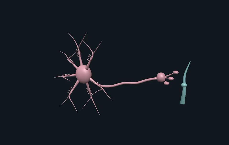
- Explain why light information starts at the retina and reaches clock-related brain circuitry through neural signals.
- Distinguish a calculated state, a laboratory phase measurement and an ordinary sleep diary.
Make a prediction
Two histories have four hours awake and two asleep. Must their final model pressure match?
- Yes, only the totals matter
- No, the order also matters
- Yes, because both last six hours
Read the explanation
From S = 0.4, wake-then-sleep ends near 0.3220; sleep-then-wake ends near 0.3967. Each interval changes the state it starts with.
Understand it
Time awake changes one part of the story
In the model, a quantity called sleep pressure rises during wake and falls during sleep. It changes smoothly; it does not become zero the instant sleep begins. Follow the solid gold curve and select one of its rising or falling sections.
Circadian timing changes another part
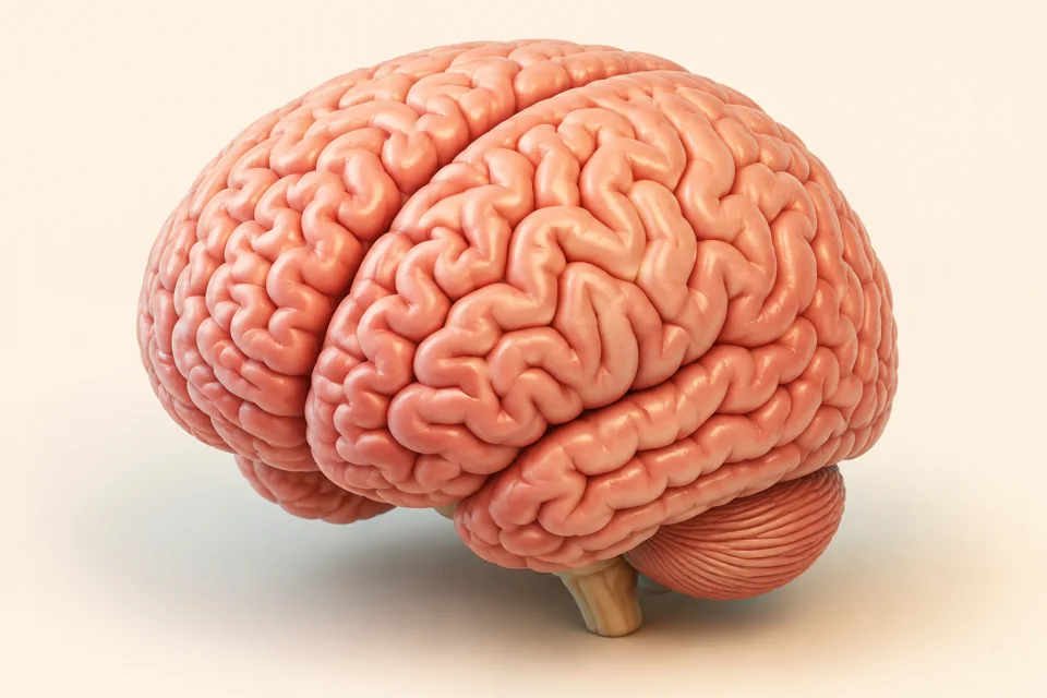
Circadian rhythms are roughly daily biological patterns. Light and dark are major timing cues, and clocks exist across many tissues. Brain circuitry including the suprachiasmatic nucleus, or SCN, helps coordinate timing. A daily pattern can continue while you are asleep.
A transition needs the appropriate boundary
Our chosen two-process model switches from wake to sleep at an upper threshold and from sleep to wake at a lower one. The daily signal moves both thresholds. Select an intersection and ask which rule was active just before the switch.
A history contains more than a total
Try four hours of modeled wake followed by two of sleep. Then reverse their order, starting at the same pressure. The totals match, but the final state differs. Each interval acts on the state left by the previous interval.
A clock change does not add elapsed time
Freeze the display, then move its local clock label from 08:00 to 14:00. No extra six hours have passed inside the model. Real adaptation to a new daily light pattern involves biology; relabeling a dial does not calculate that adaptation.
Light supplies information through the eye
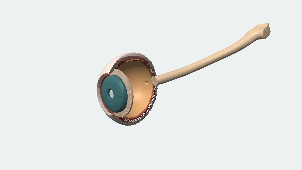
Light is detected at the retina. Specialized photosensitive retinal ganglion cells contribute signals to clock-related pathways, including projections to the SCN region. The drawing’s relay is a neural message; it is not a ray shining through the skull into a brain clock.
Look closer at the science
The declared mathematical model
We use the two-process presentation in Skeldon, Dijk & Derks (2014), Figure 1b. Wake: dS/dt = (1 − S)/18.2 h. Sleep: dS/dt = −S/4.2 h. C = sin[2π(t − α)/24 h]. The boundaries are U = 0.60 + 0.10C and L = 0.17 + 0.10C. S and the thresholds are dimensionless. The reset S = 0.17, awake at t = 0, is an authored initial condition.
What the computation does
Each branch uses its exact exponential solution. A bracketed first-crossing search switches states without resetting pressure. The plot and downloaded table sample that same trajectory. Removing circadian modulation is an intervention on this model’s structure; it is not a simulated treatment or brain lesion.
A fixed wave does not model light adaptation
The quantitative circadian signal is prescribed. It has no light-response or entrainment equation. An assumed phase shift restarts a comparison; it is not the calculated result of turning on a lamp. The anatomy and light-experiment views explain separate biological evidence.
Pressure, measurements and experience differ
EEG slow-wave activity has been used to estimate homeostatic dynamics. Parameters can vary with individuals, normalization and modeling choices. Our S is not measured EEG voltage, blood adenosine concentration, subjective sleepiness, accumulated brain damage or a health score.
Sleep has stages this model does not generate
Human sleep includes REM and three NREM stages. A binary wake/sleep trace does not tell us those stages, dream content, learning performance or a person’s exact sleep requirement. Those questions need other observations and models.
Light timing can change shift direction
Khalsa et al. studied a laboratory protocol with 6.7 hours of bright-light exposure and melatonin-phase measurements. Exposure centered before the critical phase produced delays; after it, advances. That biological phase convention is distinct from the arbitrary zero of our sine model. It is not a household-light schedule or a rule tied to one civil-clock hour.
Anatomy and hormonal output are separate
The SCN region helps coordinate timing. Clock-related signaling also influences the pineal gland, which produces melatonin. The SCN is not itself the melatonin-producing gland. Added anatomical callouts are magnified teaching locations, not measured segmented nuclei.
Where this is used
Read a model with its axes attached
An elegant curve needs a named variable, units, starting condition and rule. Keeping these visible helps distinguish a mechanism model from a sensor recording or a personal prediction.
Understand a clock mismatch
Travel can change external time labels before biological rhythms adapt. The clock comparison isolates that first distinction; it does not calculate how long adaptation will take.
Ask better questions about an experiment
What was measured: behavior, EEG, melatonin phase or a subjective report? What changed: light protocol, history or only a plotted label? Separating those questions prevents one finding from being used to claim another.
Try it yourself: A paper clock and a fictional sleep history
Supplies
- Paper and a pencil
- Three 24-hour rows, drawn or printed
- Optional tracing paper
- Fictional sleep/wake cards from this lesson
- Make a readable time row
Mark a 24-hour row. Draw one fictional 16-hour wake block and one 8-hour sleep block. These are model inputs, not recommended durations for a person.
- Add a pressure sketch
Above the row, sketch a rising wake segment and falling sleep segment. Label it model pressure, not a measured chemical level. It should stay continuous at a switch.
- Keep totals, swap order
Start a separate six-hour row at S=0.4. Compare four wake hours then two sleep hours with the reverse order. Use the calculated endpoints to check the difference; do not change your own sleep to test it.
- Move only the label layer
Keep every block fixed. Use tracing paper or another row to move civil-clock labels by six hours. Explain why the interval lengths and elapsed waking time did not change.
- Separate a diary from a measurement
If using ordinary observations, distinguish went to bed from estimated sleep began. Use a dotted mark for uncertainty and leave missing information blank. A diary does not directly measure the SCN’s phase.
- State one thing the activity cannot establish
Choose sleep stages, a personal need for sleep, or how a lamp changes phase. Explain why paper blocks and a fixed sine wave cannot answer that question. The light-study card uses a separate laboratory protocol.
Can you change clock labels without changing the length of an interval?
Use fictional cards, or record an ordinary routine afterward without changing it. No altered bedtime, alarms, caffeine, medication, light exposure or sleep duration. The record stays local and cannot diagnose a sleep condition.
Check your understanding
A fictional half-hour sleep block occurs while clock phase continues. Which modeled quantity falls because of sleep?
- Sleep pressure S
- Elapsed time
- The circadian period
Answer and explanation
Sleep pressure S The homeostatic branch changes with sleep/wake state; the prescribed circadian signal continues.
Starting at S=0.4, what remains after 4.2 hours of modeled sleep?
- Exactly zero
- About 0.1472
- Exactly 0.4
Answer and explanation
About 0.1472 One time constant leaves 1/e of the starting value. This does not establish an adequate sleep duration for a person.
An awake model has S=0.55, U=0.60 and L=0.17. Does exceeding L trigger sleep?
- Yes, either boundary works
- No, the awake state uses U
- Only if the clock labels change
Answer and explanation
No, the awake state uses U The upper boundary triggers onset while awake; the lower boundary triggers waking while asleep.
Equal wake and sleep totals in a different order must produce which result?
- Identical final S
- No possible difference
- Potentially different final S
Answer and explanation
Potentially different final S The two six-hour trials end near 0.3220 and 0.3967 from the same initial S.
At a frozen instant, local labels change from 08:00 to 14:00. What must have happened internally?
- Six hours of pressure buildup
- A six-hour biological phase advance
- Neither follows from the label change
Answer and explanation
Neither follows from the label change Elapsed time and internal reference remain unchanged by this coordinate operation.
Can the same kind of light exposure shift phase earlier or later depending on biological timing?
- Yes, in the cited laboratory protocol
- No, all light advances everyone
- Only if S is reset to zero
Answer and explanation
Yes, in the cited laboratory protocol Direction depended on phase in the cited experiment. The fixed-wave app has no light-dose response equation.
What is a displayed S=0.62?
- A blood adenosine concentration
- A measured EEG voltage
- A dimensionless model state
Answer and explanation
A dimensionless model state The app calculates a model variable and measures no bodily signal.
What can one eight-hour model sleep interval establish about a person?
- Their exact REM sequence
- Their fully restored exam performance
- Neither is calculated here
Answer and explanation
Neither is calculated here The engine tracks pressure and binary states against a prescribed circadian signal.
Sources and model limits
- A mathematical model with one published parameter set and a specified initial condition. It is not fitted to a child, adult or patient.
- S is a dimensionless state. No alertness score, diagnostic result, driving-safety judgment, exam-performance prediction or personal sleep recommendation is calculated.
- Prescribed blocks mean fictional modeled sleep or wake, not time in bed and not a guarantee that a person can fall asleep on command.
- The fixed circadian wave has no light-dose input or adaptation dynamics. Clock-label changes, assumed phase changes and elapsed time remain separate operations.
- The added daytime sleep block increases total modeled sleep. The separate order experiment holds total durations equal. These answer different comparison questions.
- The model does not simulate sleep stages, melatonin concentrations, caffeine, medication, long-term sleep-restriction effects or individual sleep requirements.
- The at-home activity observes an ordinary routine or uses supplied fictional cards. It asks for no altered sleep, alarm, caffeine, medication or light behavior.
Professional brain and eye reference illustrations
Unmodified PNGs by Servier Medical Art, CC BY 4.0. Separate schematic timing callouts are authored by Brytalearn; no exact SCN segmentation is claimed.
Servier Medical Art · BrainEye cutaway and source image reuse terms
Anterior at right, optic-nerve exit at left; the retinal lining is distinct from the orange vitreous-body context. CC BY 4.0 with attribution.
Servier Medical Art · Eye structureCircadian patterns, environmental cues and coordinated tissue clocks
Institutional fact sheet. Its fruit-fly molecular example is not presented as a complete human clock.
NIH NIGMS · Circadian RhythmsWake history, sleep need and light-related timing
Institutional sleep/wake-cycle explanation. General physiology is not an individual calibration.
NIH NHLBI · Your Sleep/Wake CycleOriginal two-process framework
Original publication record. The accessible record verifies its 1982 origin; numerical parameters here are transcribed from the later explicit mathematical presentation.
Borbély · Human Neurobiology, 1982Wake/sleep-dependent pressure with circadian thresholds
Original 1984 model abstract. Pressure need not be identified with one neurochemical substance.
Daan, Beersma & Borbély · 1984Exact equations and parameter set for this implementation
The two-process model section, equations 1–4 and Figure 1b. Other models and parameter sets in the paper are not mixed into this engine.
Skeldon, Dijk & Derks · PLOS ONE, 2014Parameter estimates depend on individual and modeling choices
Original analysis in eight healthy young men, with EEG slow-wave activity and normalization choices. These results do not define defaults for all ages.
Rusterholz, Dürr & Achermann · Sleep, 2010Direction of light-related phase resetting depends on biological timing
Original study of 21 entrained participants under a 6.7-hour laboratory light protocol, using melatonin phase. Its protocol is not a household-lamp recommendation.
Khalsa et al. · Journal of Physiology, 2003Photosensitive retinal ganglion cells project to clock-related circuitry
Original rat/mouse melanopsin-cell study. Supports a neural retinal-to-SCN route, not direct light transmission through brain tissue.
Hattar et al. · Science, 2002REM and NREM stages are distinct from the binary model
Institutional explanation of sleep stages. No stage sequence or EEG signal is generated by the two-process model.
NIH NHLBI · Sleep Phases and StagesPrescribed circadian waveforms omit light-driven oscillator dynamics
Section 2 distinguishes this limitation. Optimization schedules from the paper are not implemented.
Yin, Julius & Wen · PLOS ONE, 2021Pineal melatonin production is distinct from SCN coordination
Brain-structure descriptions, pineal-gland item. The added output callout abbreviates intervening neural signals.
NIH NINDS · Understanding SleepIndependent subject review is pending.
Read the sources and model assumptions