INTERACTIVE EXPLANATIONHow do kidneys keep useful things while making urine?
Inspect real urinary anatomy, follow a glucose marker out of blood and back again, and discover three different crossings with a ledger that never loses a marker.
Enable JavaScript to change the conditions and run the interactive experiment.
Make a discovery
The first filter does not simply recognize waste. Useful water and glucose can enter the filtrate, and selected material can return to blood later. Tubular secretion supplies another route from blood into tubular fluid. Final urine reflects all these processes, not filtration alone.
- Trace kidney → ureter → bladder → urethra without confusing blood vessels and urinary passages.
- Distinguish the glomerular capillary tuft from the surrounding capsular space.
- Explain filtration, reabsorption and secretion using both location and direction.
- Follow a useful glucose marker through filtration and recovery to blood.
- Account for selected marker identities, including transfers, storage and body exit.
- Recognize that chosen marker counts do not predict GFR, urine volume or personal health.
Make a prediction
Glucose is useful. Does that fact alone keep it out of the first filtrate?
- Yes; the glomerulus only recognizes waste
- No; glucose can be filtered and then recovered
- Yes; glucose is a red blood cell
Read the explanation
Useful does not mean unfilterable. Follow G1 across the first barrier, then through the proximal-tubule recovery route toward adjacent blood.
Understand it
Blood has two glomerular connections
Blood enters the glomerular capillaries through an afferent arteriole and leaves through an efferent arteriole. The outgoing vessel leads toward capillaries around tubules or medullary vasa recta before venous return. The efferent arteriole is not the start of the urine tube.
Filtration creates a different fluid space
Capillary pressure helps drive filtration through a layered barrier into Bowman’s capsular space. Blood cells remain vascular, while the normal barrier strongly restricts large plasma proteins. Some plasma water and small dissolved substances can cross. That includes useful substances; the filter is not a waste-recognition device.
Recover useful material through a wall
The filtrate enters the proximal tubule. Reabsorption transfers selected material from tubular fluid across epithelium and interstitium toward adjacent capillary blood. Glucose entry into proximal-tubule cells involves sodium-coupled transport proteins. Water and many solutes are also recovered, with different transport properties along the nephron.
Add selected substances by another crossing
Secretion moves selected substances from the blood side through tissue into the tubular lumen. Our C2 creatinine marker takes this route after remaining in blood at the glomerulus. C1 and C2 end on the same urine route with different histories. The displayed counts are chosen examples, not normal creatinine fractions.
Modify, collect and drain
Fluid follows proximal tubule, descending and ascending loop segments, distal and connecting tubule, then a collecting duct shared with other nephrons. Collecting structures occur in cortex as well as medulla. Final urine drains through papillary outlets, calyces, renal pelvis and ureter to the bladder.
Storage is not outside the body
The bladder stores urine before it passes through the urethra. Reaching a kidney’s drainage route, reaching the bladder and crossing the displayed outside-body boundary are three different milestones. The episode keeps stored markers in its inventory until the final step.
Look closer at the science
Filtration, recovery and secretion have different meanings
Glomerular filtration crosses from capillary blood to capsular space at the renal corpuscle. Reabsorption crosses from tubular fluid toward blood. Secretion crosses from blood toward the tubule at a tubular wall. Filtration and secretion share a broad direction but occur at different structures through different mechanisms.
Recovery needs a receiving blood route
The nephron is not an isolated tube emptying into the bladder. The efferent blood supply leads to a second capillary network near the tubules. Reabsorbed material crosses tissue into that nearby vascular route. It does not jump back upstream to the glomerular entrance.
Glucose transport has molecular machinery
SGLT proteins couple sodium and glucose entry at the proximal tubule’s luminal side. Other transport and supporting gradients complete recovery toward blood. Original studies have investigated both human transport proteins in expression systems and glucose handling in experimental animals. Their measurements are not used as human rate parameters in our marker model.
Creatinine illustrates two routes
Creatinine is a normal waste product associated with muscle metabolism. It can reach final urine through glomerular filtration and a contribution from tubular secretion. Our one filtered and one secreted marker illustrate the distinction. Their visible proportions do not describe a typical person or interpret a laboratory result.
A conserved marker cannot vanish in a wall
For a nonreacting selected substance, final urinary delivery equals filtered minus recovered plus secreted. Blood return equals the input minus that delivery after the episode empties. During playback, waiting, vascular or tubular inventory, tissue transit, storage and outside markers must also be counted. Transfer columns record events; they are not separate stockpiles.
Shape and function live at different scales
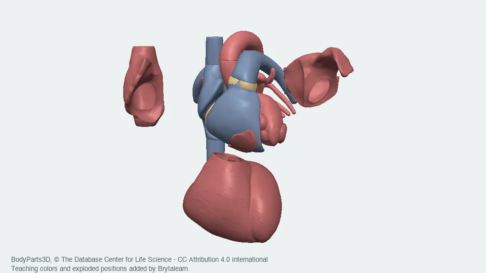
The BodyParts3D surfaces depict gross urinary anatomy. The reference illustration shows kidney regions and a nephron in context. Our glasslike route model then enlarges a nephron and its collecting duct for readability. The renal corpuscle lies in cortex; loops extend into medulla to different depths. Neither tissue transparency nor equal-size marker symbols is literal anatomy.
This covers selected jobs, not all kidney physiology
Kidneys also participate in salt and acid balance, blood-pressure regulation and hormone-related functions. Actual transport depends on many coupled solutes, gradients and regulatory signals. A detailed human-nephron computation needs far more information than nineteen visible markers; this lesson does not solve that physiology.
Where this is used
Read an anatomy illustration critically
Follow the labeled renal artery, vein and ureter in the original reference. A broad “filtered blood” label does not mean every waste molecule is gone. Source identity helps, but a clear explanation still has to state what a label means.
Understand a transport experiment
Researchers can study transport proteins in cells, or track handling in animal and human studies. Those different experimental systems answer different questions. The source list preserves those distinctions instead of treating every result as a complete human-kidney simulation.
Use the same accounting idea elsewhere
The bookkeeping skill transfers to chemical processes and water systems: specify a boundary, count what enters, track transfers and storage, then count what leaves. A correct balance helps check a model but does not prove that every biological assumption is correct.
Try it yourself: Keep track of nineteen paper travelers.
Supplies
- Four sheets of paper and a pencil
- Nineteen large paper cards or sticky notes
- Optional colored pencils
- Optional ruler or tape; cards can be torn or folded
- Draw separate lanes and a boundary
Draw a blood lane and a tubular-fluid lane. Connect them with one glomerular filtration crossing and later recovery/secretion crossings separated by a tissue strip. Add bladder storage, a urethra arrow and an outside-body box. Hands supply the movement; paper does not model pressures or membranes.
- Name every traveler once
Make RBC1–2, A1–2, W1–10, G1–2 and C1–3: nineteen cards. Use names so colors are optional. Put all cards in incoming blood and count each species. Equal-size cards do not represent equal molecular sizes or amounts.
- Filter the chosen sample
Move W1–6, G1–2 and C1 across the glomerular crossing into the tube lane. Keep the other ten cards vascular and advance them through the efferent route toward adjacent capillaries. The nine filtered cards are an authored sample, not a measured filtration fraction.
- Recover, then add by secretion
Move G1–2 and W1–5 from tubular fluid through the tissue strip to adjacent blood. Pause once in the tissue and keep counting those cards. Now move C2 the opposite way, from blood through tissue into the tubule. Label C1 “filtered” and C2 “secreted.”
- Separate return, storage and exit
Count sixteen returning-blood cards. Move W6, C1 and C2 along the drainage route into bladder storage. None is outside yet. Then move those three through the urethra arrow. Check every species: blood return plus outside cards equals its starting count.
- Replay one accounting change
Reset all nineteen IDs. Keep every rule except recover only W1–3 instead of W1–5. Predict before moving: three water cards now join two creatinine cards on the urine route; fourteen cards return in blood. Explain why this result cannot tell anyone how much water to drink or whether a kidney is healthy.
Can you follow a round trip, two routes to urine, and every marker that is still in storage?
A dry paper model only. No body fluids, samples, medicines, food, liquids or small beads. A kitchen filter cannot demonstrate selective recovery of dissolved glucose and is not a substitute for this activity.
Check your understanding
Which order carries urine from a kidney to outside the body?
- Ureter → bladder → urethra
- Renal vein → bladder → ureter
- Urethra → bladder → ureter
Answer and explanation
Ureter → bladder → urethra The ureter carries urine to storage; the urethra is the final outlet. Renal veins belong to circulation.
Our useful glucose marker appears in the first filtrate. What can happen next?
- It must leave the body because filtration is final
- It can return toward blood through tubular reabsorption
- It becomes a red blood cell
Answer and explanation
It can return toward blood through tubular reabsorption The tubule modifies the fluid after filtration. G1 crosses the wall and tissue into adjacent blood.
RBC1 passes through glomerular capillaries. Which exit continues its blood route?
- Bowman’s capsular space
- The proximal tubular lumen
- The efferent arteriole
Answer and explanation
The efferent arteriole It remains vascular and proceeds toward the second capillary network. Capsular space and tubular lumen contain a different fluid.
Which crossing is reabsorption?
- Tubular fluid → tissue → adjacent blood
- Blood → tissue → tubular fluid
- Glomerular blood → capsular space
Answer and explanation
Tubular fluid → tissue → adjacent blood Recovery points toward blood at a tubular wall. The opposite tubular crossing is secretion; the glomerular crossing is filtration.
C2 remained in blood at the glomerulus and entered the tubule later. What added it?
- Recovery from final urine
- Tubular secretion
- Filtration through the ureter
Answer and explanation
Tubular secretion Its recorded history shows the blood-to-tubule crossing through tissue. C1 reached the same destination by filtration at a different structure.
Six W markers are filtered, five recovered and none secreted. How many reach the final urinary route?
Answer and explanation
One 6 − 5 + 0 = 1. Recovery transfers five existing markers toward blood; it does not create five extra markers.
Three final markers are in the bladder before the final exit step. How many have crossed the displayed outside-body boundary?
Answer and explanation
Zero Storage remains within the displayed boundary. Follow the urethra step to move them outside.
Recovering five W markers instead of three establishes which result?
- A measured healthy GFR
- Fewer W markers remain on this chosen urine route
- A personal daily drinking recommendation
Answer and explanation
Fewer W markers remain on this chosen urine route The conclusion follows from the chosen ledger. There are no physiological volumes, flow rates or personal measurements from which to infer the other claims.
Sources and model limits
- Nineteen selected markers with authored transfer rules. Counts are not molecule-size ratios, concentrations, masses, volumes, probabilities, measured fractions or physiological rates.
- This closed episode omits marker production, consumption and many exchanges. Water-recovery comparisons are two chosen inventories, not hydration states or kidney-efficiency controls.
- The two albumin markers remain vascular as a coarse representation of strong restriction; real trace protein handling is below this model’s resolution. Blood cells have no tubular route in this example.
- The original anatomical atlas is an adult male reference, not a patient scan or universal anatomy. Only selected renal vessel segments are included.
- The nephron, barrier and tubule-wall scenes are original schematic geometry. They preserve named route relationships, not measured dimensions, transparent tissues, permeability, pressures or single-molecule trajectories.
- Marker movement through tissue does not simulate channels, coupled-ion transport, osmosis, energy use or hormonal regulation. Glucose and water recovery here do not stand for all segment-specific physiology.
- No GFR, urine volume, creatinine concentration, medication response, disease severity, health score, diagnostic result or drinking recommendation is calculated.
- The dry paper activity has not been classroom-trialed. Independent subject review, browser/device rendering, accessible interaction review and encoded-video checks remain pending.
Glomerular filtration and tubular recovery have distinct roles; kidneys also perform other regulatory functions.
Institutional physiology explanation. Its broad daily-filtering language is not used as a GFR parameter.
NIDDK · Kidney functionKidneys, ureters, bladder and urethra form distinct stages of urine production, transport, storage and exit.
Supports the visible anatomical route. No hydration or self-testing guidance is added.
NIDDK · Urinary tractThe glomerular and peritubular vascular routes coexist with separate nephron lumens and tissue regions.
Institutional histology guide supports both arterioles, receiving capillaries, cortex and medulla. Source microscopy is linked, not redistributed.
SIU School of Medicine · Kidney histologyRenal corpuscles and tubular segments occupy different regions and connect to collecting structures.
Supports the nephron-and-collecting-duct organization. One illustrative loop depth does not represent every nephron.
University of Leeds · NephronCapsular space, vascular and urinary poles, tubule segments and collecting drainage are distinct structures.
Original human H&E specimen reference identifies the University of Minnesota department. Its microscopy image is not copied into the lesson.
Human kidney slide · Histology GuideEarly proximal glucose recovery involves SGLT2 transport machinery.
Original mouse experiments, including knockout and micropuncture. Species and experimental context are retained; reported percentages are not human model parameters.
Vallon et al. 2011 · Glucose recoveryHuman renal SGLT1 and SGLT2 proteins couple sodium and glucose transport.
Original expression-system experiments in HEK293T cells. Supports the molecular mechanism, not a whole-person kidney prediction.
Hummel et al. 2011 · Human glucose transportersCreatinine secretion is distinguishable from filtration and urinary excretion.
Original 1996 MDRD study, JASN 7:556–566. Its renal-disease population and rates are not used to describe normal proportions in this toy ledger.
NIDDK Repository · Creatinine handling studyQuantitative nephron transport requires conservation and many segment-specific, coupled relationships.
Original computational research. Its equations and parameters are not implemented here; it supports the boundary between marker bookkeeping and physiology simulation.
Layton & Layton 2019 · Human-nephron modelCapillary pressure and a layered endothelial, basement-membrane and podocyte barrier contribute to filtration.
Only qualitative layer and mechanism claims are used. No sharp molecular-size cutoff or suspect unit value is imported.
University of Leeds · Renal corpuscleCreatinine is a normal waste product; a toy marker cannot interpret a laboratory result.
NLM-authored explainer supports marker identity. No thresholds, testing instructions or clinical interpretation are provided.
MedlinePlus · CreatinineOriginal source surfaces identify the two kidneys, ureters, bladder, urethra and selected renal vessels.
Version 4.0 OBJ source names and concept IDs are preserved in the local urinary manifest. One common transform retains their relative positions.
BodyParts3D · Original atlasThe source archive’s current notice specifies CC Attribution 4.0 International.
BodyParts3D, © The Database Center for Life Science. Older OBJ headers retain former wording; the distribution follows the current archive notice.
BodyParts3D · Current licenseThe credited institutional reference illustrates a kidney section and enlarged nephron.
Original labeled image, unchanged. Credit: National Institute of Diabetes and Digestive and Kidney Diseases, National Institutes of Health.
NIDDK · Kidney and nephron imageNIDDK-produced reusable content requires source acknowledgment and must not imply endorsement.
The specific media entry and policy support the reference image’s use. Third-party exceptions elsewhere on the site are not assumed reusable.
NIDDK · Reuse policyIndependent subject review is pending.
Read the sources and model assumptions