INTERACTIVE EXPLANATIONHow do your inner ears help you balance?
Brake a virtual chair, open a real inner-ear reconstruction, and solve a motion-clue puzzle. Discover why actual movement, sensory responses and what you feel are different.
Enable JavaScript to change the conditions and run the interactive experiment.
Make a discovery
Stopping a movement does not instantly settle every sensory response. Different kinds of clues help the brain interpret motion.
- Locate the canal loops and neighboring cochlea in real anatomical evidence.
- Distinguish actual head rotation from a dynamic mechanical response and a feeling.
- Explain starting, steady rotation, braking and relaxation in a bounded model.
- Show how two different motions can produce the same declared gravity-and-acceleration vector.
- Compare complementary visual, vestibular and body/support information.
- Preserve the difference between observed anatomy, calculated values and illustrative motion.
Make a prediction
The chair has stopped, but the model response is negative. What does that mean?
- The chair secretly turns backward.
- A mechanical response can remain after motion stops.
- Every person feels this exact amount of dizziness.
Read the explanation
The actual speed and model state are different. Interpretation and feeling are additional processes.
Understand it
Make something happen first.
Start the virtual rider, let the chair turn, then brake. The chair and rider move together. You can remain comfortably still, or use the still-stage cards. Look at the two readouts: actual chair speed and a simplified mechanical response inside the ear.
The chair is still. A response remains.
In this example, the canal response changes sign during braking and takes time to settle afterward. That does not make the chair secretly turn backward. A sensory response and the motion that caused it are different quantities. The brain’s interpretation is another step.
Meet the semicircular canals.
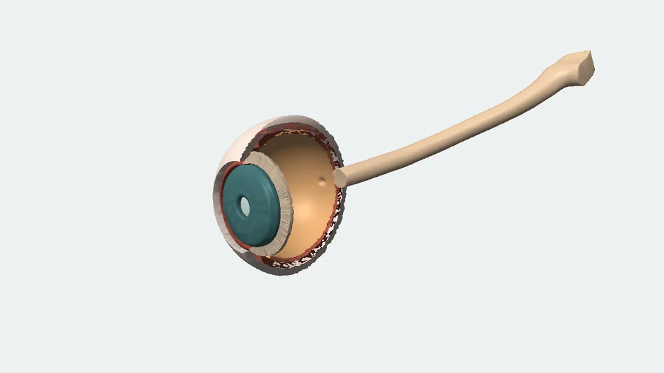
Inside each inner ear are small membranous ducts, housed in bony channels. Fluid mechanics acts on a flexible structure called the cupula in each widened ampulla. Its motion couples to sensory hair cells. The real 3D scan shows bony space; our enlarged moving cupula is a separate teaching illustration.
Another clue cannot name its own cause.
The utricle and saccule are the otolith organs. Their sensory tissues couple to a layer containing tiny dense calcium-carbonate particles called otoconia. They respond to the combined effects of gravity and linear acceleration. Our two virtual cabins show why that combined clue alone can fit more than one motion.
Put different clues together.
The eyes provide information relative to a visual scene. Muscles, joints and contact provide information about the body and its support. Inner-ear signals add information about motion. A scene or support may itself move, so these are complementary reference frames—not identical votes with fixed percentages.
Give the idea its name.
Vestibular means relating to the inner ear’s balance-and-motion system. Post-rotation motion sensations involve dynamic signals and their interpretation. This lesson explains one relevant mechanism; dizziness can have many causes, and this model does not assess an individual person.
Look closer at the science
A sensor with a history
The selected single-mode reduction is dy/dt + y/τ = α, with angular acceleration α, example time constant τ = 4 s and scaled state y. The plotted index is y divided by π/3 rad/s. Finite ramps avoid treating a speed discontinuity as an ordinary time step. Natural canal, afferent and central responses are more complex than this mechanical reduction.
Why the response changes during a constant speed
During a constant-speed interval, angular acceleration is zero and the model state relaxes exponentially. During braking, the acceleration drives it the opposite way. A response can therefore cross zero before physical rotation stops. With no further input, one time constant reduces its magnitude to 1/e, about 0.368, rather than erasing it.
The worked example
A 2 s start reaches 60°/s; an 8 s cruise precedes a 2 s stop. The normalized response is approximately +0.787 at the end of the start, +0.107 before braking, −0.722 when motion stops, and −0.266 four seconds later. Keeping the same cruise and braking over 8 s instead gives approximately −0.418 at the stop. These are calculated model states, not measured feelings.
A material uncertainty
The 2016 geometry-based study reports a human lateral-duct model time constant around 4.0 ± 0.8 s. An earlier indirect eye-response fit reports 4.2 ± 0.6 s in ten humans. Other biomechanical estimates report about 16–20 s under different assumptions. We do not resolve this disagreement or claim four seconds is a universal human constant. The 2016 uncertainty is not a between-person standard deviation.
The same two-axis vector
In our head-fixed right/down convention, f = g − a. A stationary 15° tilted cabin has f = [2.538148, 9.472497] m/s². An upright cabin can match it with acceleration [−2.538148, 0.334153] m/s²: leftward and slightly downward. Horizontal acceleration alone matches only one component. These momentary constructed vectors do not make the entire sensory histories identical.
What the real geometry establishes
The Wimmer dataset contains co-registered imaging and bony-labyrinth reconstructions from 23 specimens. Our rotatable F01 is a left-side micro-CT surface with 87,155 vertices and 174,318 triangles. Its original sampling is 0.06 mm; the largest source bounding-box extent is about 16.264 mm. No membrane, cupula mechanics or head-aligned yaw frame is inferred from that bony surface.
Anatomy is not physiology
David et al.’s stained micro-CT shows smaller membranous structures inside bony space. Direct toadfish measurements in a separate study distinguish cupula motion from diverse afferent responses; those fish times are not human constants. Human and primate motion studies test additional neural and sensory-integration processes. Our anatomy, mechanical model and cue cards keep these evidence roles distinct.
Where this is used
Keeping a view steady
Vestibular information contributes to gaze stabilization during everyday head movement. It operates with neural processing and eye movements beyond this one-state model; a canal response is not a finished command for an eye.
Reading a ride’s motion
A familiar start, cruise and stop supplies an accessible way to compare physical motion with a dynamic sensor. The virtual rider’s finite profiles are authored examples, not instructions for operating a ride or a person.
Investigating several kinds of evidence
Researchers and clinicians may compare anatomy, motion, eye responses and other information because no one measurement establishes every part of balance. This learning activity makes no diagnosis or personalized assessment.
Try it yourself: A seated motion detective
Supplies
- One sheet of paper or the screen worksheet
- Pencil
- Three small paper markers
- Separate three kinds of information
Make columns for actual motion, canal model and other information. Use the supplied end-of-start, end-of-cruise, stop and four-seconds-later records.
- Predict before revealing
Place forward, zero or reverse markers for actual speed and model response. Then check the numbers. At the stop, actual motion is zero while the response remains negative.
- Compare one changed brake
Keep the 8-second cruise. Compare stopping over 2 seconds and over 8 seconds: approximately −0.722 versus −0.418 at the respective stops. Write what changed and what stayed fixed.
- Solve the ambiguous clue
Place the tilted and accelerating cabin cards beside their matching two-axis vector. Choose an extra clue—recent rotation, fixed outside scene or body/support relation—and explain its possible use.
- Check actual anatomical evidence
In the full published figure, human anatomy is row a. Find blue bony space and the smaller red membrane. Finish: “The image shows ___. The simulation adds ___.”
Can you explain a movement from its clues without moving yourself?
Use the supplied records while seated. This is a paper interpretation activity, not a spinning experiment, balance test or personal health assessment.
Check your understanding
The chair has just stopped. Its model response is still negative. What follows?
- The chair secretly turns backward.
- A mechanical response can persist after rotation stops.
- Every person feels this exact dizziness.
Answer and explanation
A mechanical response can persist after rotation stops. The state relaxes after the physical input ends.
Our response gets smaller at a constant turning speed. What does that show?
- The canals are useless after the start.
- These are dynamic sensors, not perfect constant-speed meters.
- The model has switched to hearing.
Answer and explanation
These are dynamic sensors, not perfect constant-speed meters. Response depends on the time course of movement.
What couples canal fluid mechanics to sensory hair bundles?
- A flexible cupula.
- Water drops passing through a hole.
- The bony loop stretching like rubber.
Answer and explanation
A flexible cupula. The cupula is a compliant structure spanning the widened duct.
What influences the otolith organs’ mechanical input?
- Head direction alone.
- Constant traveling speed alone.
- Gravity and linear acceleration.
Answer and explanation
Gravity and linear acceleration. The combined input does not separately label its two causes.
Two cabins match the complete two-axis vector. What is established?
- Their motions must be identical.
- That vector alone cannot choose between these cases.
- All their body and visual histories match.
Answer and explanation
That vector alone cannot choose between these cases. A shared output can have more than one physical cause.
How should we describe vision, body and inner-ear information?
- Vision always wins.
- Each always contributes a fixed percentage.
- They supply complementary clues with different references.
Answer and explanation
They supply complementary clues with different references. The usefulness of a reference depends on its context.
What is the actual 3D reconstruction?
- One left human bony labyrinth from imaging.
- A movie of fluid in a living child.
- Everyone’s exact average inner-ear shape.
Answer and explanation
One left human bony labyrinth from imaging. Its static anatomical provenance is visible and downloadable.
What does τ = 4 seconds mean in this example?
- Every dizzy spell ends in four seconds.
- An unforced model response becomes 1/e as large in four seconds.
- The response becomes exactly zero in four seconds.
Answer and explanation
An unforced model response becomes 1/e as large in four seconds. An exponential time scale is not a symptom duration.
Sources and model limits
- The dimensionless response index is not perceived speed, percentage dizziness, a measured cupula displacement, a nerve firing rate or a balance score.
- Four seconds is an explicitly chosen example time constant, not a universal human physiological constant or the duration of a dizzy spell. Published estimates depend on assumptions and remain disputed.
- The one-mode reduction omits the fast mechanical mode, hair-cell/afferent processing, bilateral canal coupling, central velocity storage and complete postural or perceptual integration. It is not applied to impacts, sound or high-frequency vibration.
- F01 is one static ex-vivo left bony-space reconstruction. It is neither a live fluid movie nor an average person. Its source coordinate frame is cochlear, not a measured head-yaw sensitivity frame.
- The animated cupula is an enlarged, original signed illustration. It spans the ampulla; no fluid is shown completing laps through a hole. Its dimensions and deflection are not measured from F01.
- The cabin puzzle matches a declared instantaneous two-axis mechanical vector. It does not match all sensory histories or calculate the brain’s interpretation. No fixed sensory-weight percentages are assigned.
- The camera and room do not rotate with the rider. The paper activity uses supplied records while seated; it is not an induced-spinning experiment or vestibular-function test.
Vestibular anatomy and complementary sensory information.
Agency-owned descriptions of canals, cupula, hair cells, otolith organs and multiple causes of dizziness. No outdated prevalence statistic or clinical maneuver is imported.
NIDCD · Balance DisordersSmaller membranous anatomy and geometry-dependent mechanical modeling.
Scientific Reports 6, 32772. Full Figures 1 and 2, captions, methods and Table 2 support the anatomy distinction and declared four-second example. Figures CC BY 4.0; human panels/specimen differences remain labeled.
David et al. (2016) · original micro-CT and mechanics paperIndirect peripheral and central mode fitting must be distinguished.
Fitted mean 4.2 ± 0.6 s in ten humans from slow-phase eye-velocity responses. Verified abstract only; no unverified methods or human symptom predictions.
Dai et al. (1999) · original study abstractMechanical displacement and neural response can have different dynamics.
Original simultaneous optical and afferent study in toadfish. Fish parameters are not human constants. The paper is linked; noncommercial-licensed figures are not redistributed.
Rabbitt et al. (2009) · direct cupula and nerve measurementsCanal information can contribute to interpreting tilt and translation.
Five rhesus monkeys and eye-movement outcomes under controlled motions; intervention in two animals. Supports the interpretation problem, not a direct report of a human feeling.
Angelaki et al. (1999) · original tilt/translation studyGravity and linear acceleration pose an interpretation problem.
Human eye responses tested against internal-model predictions. Our vector construction illustrates the mechanics ambiguity without reproducing provocative laboratory motions.
Merfeld, Zupan & Peterka (1999) · human motion studyUse of sensory orientation information depends on conditions.
Controlled visual-surround and support-surface experiments. No fixed universal percentages, personalized weights or home balance protocol are derived.
Peterka (2002) · human sensory integrationImaging and bony-labyrinth surface provenance.
Co-registered clinical CT/micro-CT and segmented surfaces from 23 human specimens. F01’s bony space is separate from David’s soft-tissue specimens and this mechanical model.
Wimmer et al. (2019) · original data descriptorActual licensed left-side 3D anatomy.
Version 1.0, CC BY 4.0. F01 descriptor confirms LEFT. Exact topology retained; centered, converted from millimeters to meters and recolored. Source and derivative hashes are in the downloadable provenance record.
Wimmer et al. · original dataset, F01Material disagreement in human time-scale estimates.
Used specifically to disclose the approximately 16–20 s estimate and its assumptions. It does not override the original studies or settle a universal constant.
Rabbitt (2019) · source-owned biomechanical reviewIndependent subject review is pending.
Read the sources and model assumptions